
Education
Brief History of X-ray CT
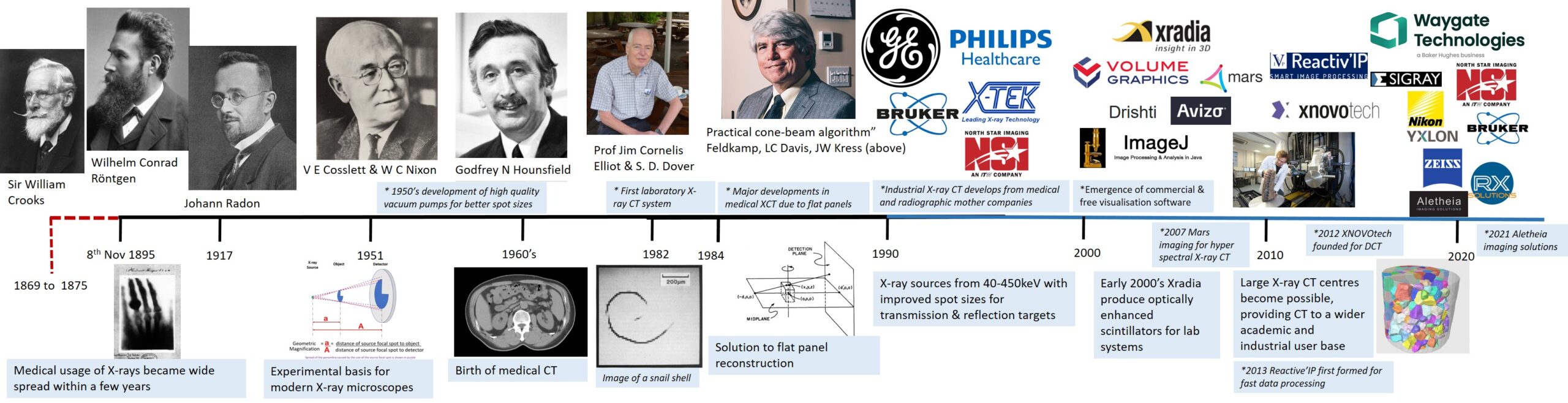
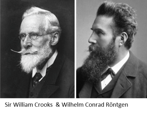
Wilhelm Conrad Röntgen (1845-1923) discovered X-rays in 1895 and was, in most respects, a foundational figure in what would become radiology. Wilhelm discovered the effect of X-rays to create an image on a fluorescent screen and photographic plates of the day from an X-ray source, previously “unknown radiation”, while experimenting with electrical discharge tubes. Then he observed fluorescence and subsequently demonstrated that this radiation could expose photographic plates and penetrate materials. It is important to mention that the X-ray source used by Wilhelm was likely a Crookes tube (or similar device) which was invented by Sir William Crookes (1832-1919) an English chemist and physicist at the royal College of Chemistry. Crookes developed experimental vacuum and electrical discharge tubes and in 1875 observed cathode rays being generated.
Wilhelm published his discovery of X-rays under the title “On a new kind of rays”, on the 28th December 1895, where “X” was the unknown radiation of the time. His work on X-rays resulted in him being awarded the first Nobel Prize for physics in 1901. It can be noted that Wilhelm never patented his work and quickly realised the benefits X-rays to developing medical science at the time and non-invasive investigations of the body. The use of X-rays were quickly adopted within medicine and were used in the diagnosis of soldiers in WW1 to find bullets, shrapnel and fractures.
- Images shown of Sir William Crooks & Wilhelm Conrad Röntgen are sourced from William Crookes – Wikipedia & Wilhelm Röntgen – Wikipedia
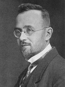
In 1917 the Austrian mathematician Johann Radon (1887-1956) provided the formula for the inverse transform. This is an important concept that would be applied almost 40 years later with the birth of medical X-ray CT. His work demonstrated that a two-dimensional function could be reconstructed from its line integrals. In simple terms this explains how 3D structural information from an X-ray passing through an object is transformed to 1D data which can be used to form a sonogram from the radiographic information as the object is rotated over 360 degrees. This proved an essential mathematical foundation for computed tomography, in which X-ray attenuation measurements acquired from multiple angles are used to reconstruct cross-sectional images of an object.
- Image shown of Johann Radon was sourced from Johann Radon – Wikipedia
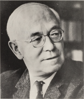
In 1951, V.E. Cosslett (1908–1990) & W.C. Nixon invented the X-ray shadow microscope at the University of Cambridge. They showed that moving an object closer to the focal spot of an X-ray source, when there is a fixed detector position, creates an increasingly magnified (shadow) image of the object at the detector. This is the fundamental science behind magnification in modern X-ray instruments. They also described the penumbra (Focal spot blur) effect that results due to the combination of the X-ray focal spot size, imaging geometry (magnification setup) and the X-ray detector pixel pitch. At high magnifications when the X-ray focal spot size approaches the X-ray detector pixel pitch the feature edge blurring can span across the pixel boundaries resulting in a blurred image.
- Image shown of V E Cosslett was sourced from Vernon Ellis Cosslett, 16 June 1980 – 21 November 1990

In 1979, Sir Godfrey Newbold Hounsfield (1919 –2004) and Allan MacLeod Cormack shared the Nobel Prize for Physiology or Medicine. Hounsfield was an English electrical engineer developed the diagnostic technique of X-ray computed tomography (CT). They developed the first clinically successful X-ray computed tomography scanner with the first patient scanned at Atkinson Morley’s Hospital in London on 1 October 1971. His name is now immortalised in the Hounsfield scale which is a quantitative measure of radiodensity used in evaluating CT scans. This can be considered the birth of medical X-ray CT which is now a standard instrument found throughout hospitals around the world.
- Image shown of Sir Godfrey Houndsfield was sourced from Godfrey Hounsfield – Wikipedia

The next major step was the development of micro tomography (micro CT) systems. This work was pioneered by Professor Jim Cornelis Elliott at the London Hospital Medical College and David Dover from Kings College London who invented and built the first micro CT system in the early 1980’s. This work was published in 1982 in the Journal of Microscopy and was titled “X-ray micro tomography”. This research represented a great leap in X-ray CT spatial resolution from the millimetre range to the 15 to 50 micrometre range. The resolution capability was originally demonstrated using a snail shell as shown in the timeline above. This design of a static X-ray source and detector with specimen placed on a rotating stage is now the standard design concept used in both Research and industrial X-ray CT systems.

The next major change occurred in 1984 when L A Feldkamp, LC Davis, JW Kress (FDK) published their reconstruction solution to cone beam X-ray CT using a flat panel. Previously X-ray CT was a slow process since only a line could be reconstructed at a time. However the FDK algorithm approximated the filtered back projection over an entire panel rather than a single line. This meant that whole sections could be analysed with a single scan resulting in a significant decrease in both data collection and reconstruction time. The FDK algorithm is now used throughout the X-ray CT industry.
- Image shown of L A Feldkamp was sourced from Who We Are — KressWorks Institute
The late 1980’s and early 1990’s saw the rapid development of X-ray CT with major companies such as GE and Philips Health care within the medical sector and the development of industry-based instruments by X-TEK and North Star Imaging (NSI).
With the expansion of X-ray CT instruments there was a growing need for data analysis which came in two forms. VGSTUDIO was developed in 1997 by Volume Graphics for 3D CT slice image stacks and in the same year ImageJ was by Wayne Rasband at the National Institutes of Health. Today ImageJ is a free software used by both academics and industry across the globe while VGSTUDIO is predominantly used for industrial applications due to its strong GD&T tools for component verification.
Founded In 2000 XRADIA produced optically enhanced scintillators for laboratory X-ray CT systems resulting in sub-micron imaging capabilities. XRADIA had a strong background in X-ray optics due to their experience with synchrotron beamline optics. This experience with synchrotron beamlines influenced their laboratory X-ray microscopes. These systems use small X-ray detector panels, where the X-ray photons create visible light when they interact with the detector material, which is then attached to optical lens to increase the magnification. Since these systems rely more on the optical magnification factor rather than the geometric magnification a much shorter FDD is required. This results in an improved image contrast for low attenuating materials. The combination of high magnification coupled with high sensitivity resulted in their widespread success in imaging difficult low attenuating materials such as carbon fibre and biomaterials.
In the early 2000’s laboratory X-ray CT systems were starting to appear in greater numbers and by 2010 a number of X-ray facilities had been established. Within the UK X-ray CT dedicated facilities such as University of Southampton’s imaging laboratory was opened in 2007 (later renamed µ-vis) which was followed by The Henry Mosely facility (2008) at Manchester, both of which supported a number of X-ray CT systems covering a range of keV and spatial resolution capabilities for research and industry applications.

Other notable developments in X-ray CT was the introduction of AVIZO in 2007 which offered a unique analysis platform for X-ray CT data analysis and research which became popular in academic institutions due to its research flexibility and image processing capabilities. In 2012 XNOVOtech was founded for diffraction contrast tomography. This technology uniquely allows the diffraction spots from a poly chromatic X-ray source to be used in order to map the grain structure of materials in 3D. This technology has been applied widely in the ZEISS XRADIA X-ray Microscope systems. In 2013 IPSDK was released by Reactiv’IP, The software was developed as a data analysis driven package resulting in rapid analysis with a transparent analysis methodology. Finally in 2016 ORS (Object Research Systems) developed the Dragonfly software. This software has strong visualisation capabilities combined with data analysis and has become very popular with in academia.
Today X-ray CT is a diverse industry with companies offering multiple platforms for both industrial and academic activities. Laboratory systems have spatial resolutions varying from the mm range for high powered LINAC sources down to the sub 100nm range for instruments from ZEISS XRADIA and SIGRAY. The large industrial X-ray CT companies such as Waygate Technologies and COMET YXLON continue to push the boundaries of industrial X-ray CT with multi high powered sources with smaller X-ray spot sizes for improved resolution while their smaller systems push spatial resolution capabilities towards the 500nm range. Newer companies such as RX Solutions and BRUKER offer new capabilities that are impacting in both academia and industry. In the area of data analysis software is now widely available with companies occupying niche solutions. The growing CT community has also had a substantial impact in the free data analysis software area with platforms such as ImageJ having a wide user base that adds to its capabilities.